Adventure in Australia

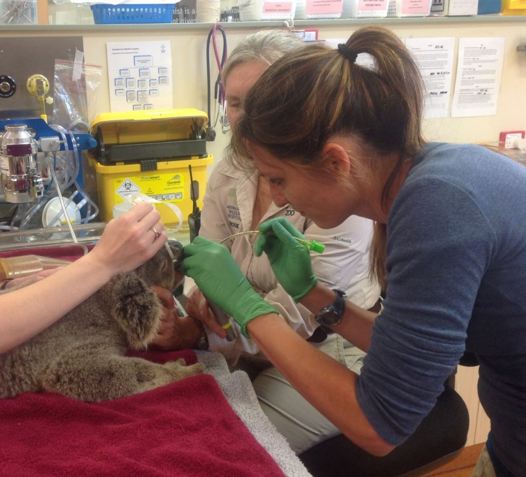
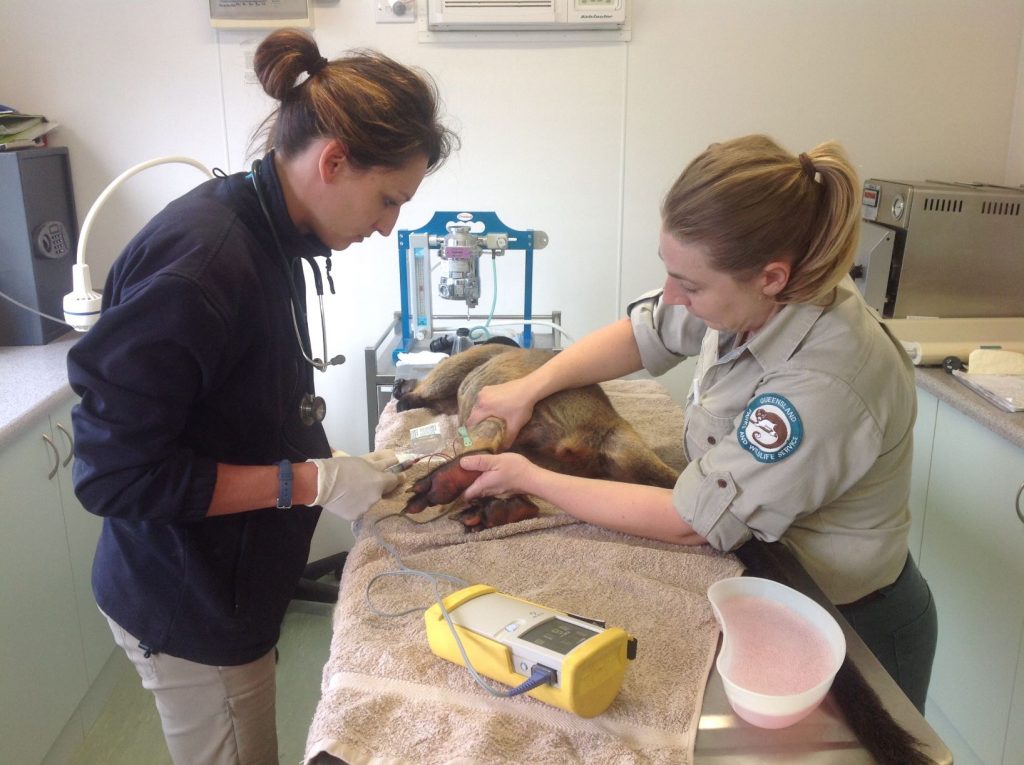
In July 2016, I went to Australia for a couple of weeks; it was one of the most incredible adventure I had during my residency. The East Coast is very much European in matter of food and even culturally. My mentor was Dr. Blyde and I spent time at Sea World Australia (doing aquatic medicine), Australia Zoo (Wildlife rehabilitation Center) and Fleay Wildlife Park. We were extremely busy.
One of my best adventures and memories. I met wonderful human beings, dedicated to the animals. I learnt one motto from my dear mentor, that will follow me from now on » Good work with good people« . Imagine taking off Florida to LA and then get ready for a 15 hours-flight! to Brisbane. I rented a manual car (do not forget to drive on the British side of the road!) at 6am and then I had to drive one hour South of Brisbane. I was so jet-lagged! I could not believe I was in Australia!
As soon as I arrived, I was sat right away into a long-nosed fur seals (Arctocephalus forstei) disease outbreak (check this paper out). These tiny pups were very sick and we were treating and rehabilitating them, running a bunch of diagnostic tests.
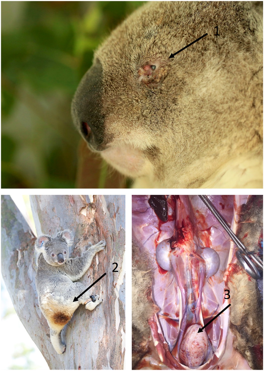
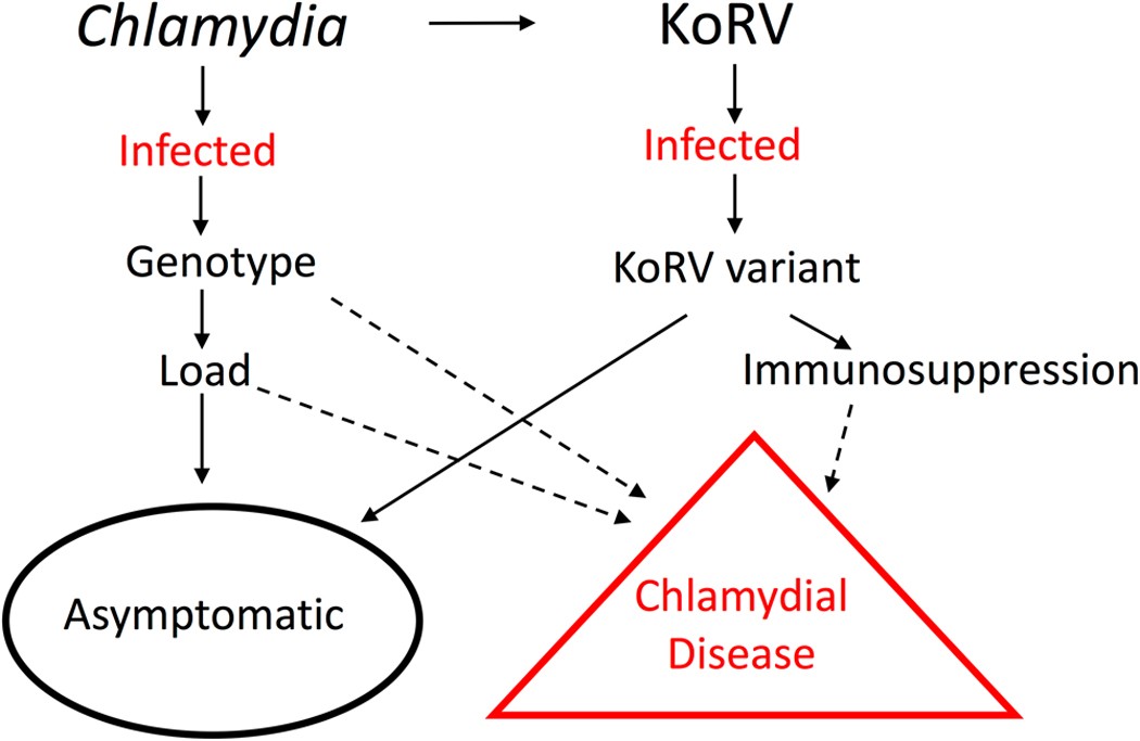
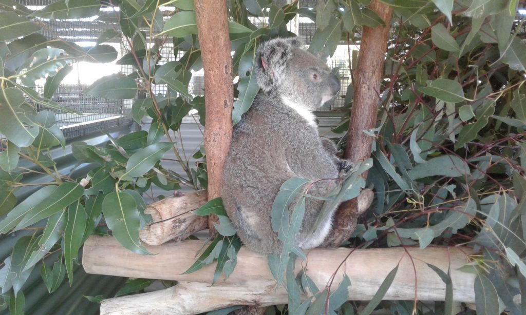
Then we went to Australia Zoo Wildlife Hospital, which has a wonderful hospital for local wildlife. They have a huge rehabilitation space dedicated to koalas with a space for healthy, ready to be released animal and another one for sick animals. Apart from deforestation (encroachment) due to human activity and more recently devastating fires, and trauma (hit by car, dog attacks); koalas are dealing with infectious diseases. Two diseases are of concern, koala retrovirus (KoRV which is a gammaretrovirus) and Chlamydia (bacteria) infection. Koala retrovirus has a prevalence of 100% in the North East Australia. There are 3 variants of this virus (A, B and J). The B variant is associated with leukemia and lymphosarcomas. Chlamydiosis in koalas is due to 2 bacteria Chlamydia pneumoniae (biovar Koala) and Chlamydia pecorum also found in cattle, sheep and pigs. Chlamydiosis can be asymptomatic (inapparent disease only discover if doing ultrasound or during post-mortem examination) or symptomatic (keratoconjonctivitis, cystitis what is called « dirty tail », infertility and rhinitis/ pneumonia). There is evidence in the literature that chlamydiosis is associated with KoRV-2 infection. The Center has set up a triage protocol to better characterize the status of each animal coming into rehabilitation and to decide if treatment is appropriate or not.
Check these papers out:
KOALA RETROVIRUS: A REVIEW
Matthew E Kinney, Geoffrey W Pye. J Zoo Wildl Med. 2016 Jun;47(2):387-96.
Koala retrovirus (KoRV) is a gammaretrovirus that has been identified in both captive and free-ranging koalas ( Phascolarctos cinereus ) with variable geographic distribution in Australia. KoRV is capable of both exogenous and endogenous transmission, which provides an interesting research platform for scientists to study active retrovirus endogenization into a host genome and offers veterinary scientists an opportunity to examine the clinical consequences of KoRV infection in koalas. Causation between KoRV and frequently recognized clinical conditions associated with immune suppression and neoplasia in koalas has not been definitively established, however research continues to evaluate a potential association. Three KoRV variants, KoRV-A, KoRV-B, and KoRV-J, have been the most thoroughly described and preliminary evidence suggests KoRV variability may be fundamental in host pathogenicity. In addition to reviewing what is currently known about KoRV, this article discusses treatment, management, and future research directions.
The prevalence and clinical significance of Chlamydia infection in island and mainland populations of Victorian koalas (Phascolarctos cinereus)
Chlamydia infection is known to impact the health of koalas (Phascolarctos cinereus) in New South Wales (NSW) and Queensland, but the clinical significance of Chlamydia infections in Victorian koalas is not well described. We examined the prevalence of Chlamydia infection and assessed associated health parameters in two Victorian koala populations known to be Chlamydia positive. The same testing regimen was applied to a third Victorian population in which Chlamydia had not been detected. We examined 288 koalas and collected samples from the urogenital sinus and conjunctival sacs. Detection and differentiation of Chlamydia species utilized real-time PCR and high-resolution melting curve analysis. Chlamydia pecorum was detected in two populations (prevalences: 25% and 41%, respectively) but only from urogenital sinus swabs. Chlamydia was not detected in the third population. Chlamydia pneumoniae was not detected. Chlamydia pecorum infection was positively associated with wet bottom (indicating chronic urinary tract disease) in one Chlamydia-positive population and with abnormal urogenital ultrasound findings in the other Chlamydia-positive population. The prevalence of wet bottom was similar in all populations (including the Chlamydia-free population), suggesting there is another significant cause (or causes) of wet bottom in Victorian koalas. Ocular disease was not observed. This is the largest study of Chlamydia infection in Victorian koalas, and the results suggest the potential for epidemiologic differences related to Chlamydia infections between Victorian koalas and koalas in Queensland and NSW and also between geographically distinct Victorian populations. Further studies to investigate the genotypes of C. pecorum present in Victorian koalas and to identify additional causes of wet bottom in koalas are indicated.
THE PARADOX OF EUTHANIZING KOALAS (PHASCOLARCTOS CINEREUS) TO SAVE POPULATIONS FROM ELIMINATION
David P Wilson 1, Andrew P Craig 1, Jon Hanger 2, Peter Timms. J Wildl Dis. 2015 ;51(4):833-42.
Koala (Phascolarctos cinereus) populations in the wild are in sharp decline in Australia due to deforestation, road accidents, dog attacks, and disease from infection with sexually transmitted Chlamydia spp. Severely diseased koalas that are captured are euthanized for humane reasons because antibiotics are not effective. Paradoxically, we propose that euthanizing more koalas could help to increase koala population numbers. We investigated the potential impact of systematically euthanizing diseased koalas. Using data from a well-studied koala population, and an individual-based computer simulation model, we predict that such a program would result in a larger population of koalas after 7 yr than would exist without the program. If terminally diseased and sterile koalas are euthanized and other infected captured koalas are given antibiotics, chlamydial infection could be eliminated and population growth observed after 4 yr. The practical implementation of such a program would be facilitated with further development of tools to diagnose infection and internal disease in the field.
Altered immune parameters associated with Koala Retrovirus (KoRV) and Chlamydial infection in free ranging Victorian koalas (Phascolarctos cinereus)
Koala Retrovirus (KoRV) has been widely speculated to cause immune suppression in koalas (Phascolarctos cinereus) and to underlie the koala’s susceptibility to infectious disease, however evidence for immunomodulation is limited. The aim of this study is to determine whether immunophenotypic changes are associated with KoRV infection in free ranging Victorian koalas. qPCR was used to examine mRNA expression for Th1 (IFNγ), Th2-promoting (IL6, IL10) and Th17 (IL17A) cytokines, along with CD4 and CD8 in whole blood of koalas (n = 74) from Mt Eccles and Raymond Island in Victoria, Australia, with and without natural chlamydial infection. KoRV positive koalas had significantly lower levels of IL17A (p`0.023) and IFNγ (p = 0.044) gene expression along with a decreased CD4:CD8 gene expression ratio (p = 0.025) compared to negative koalas. No effect of chlamydial infection or combined effect of KoRV and chlamydial infection was detected in these populations. The decreased expression of IFNγ could make KoRV infected koalas more susceptible to persistent chlamydial infection, and a decrease in IL17A could make them more susceptible to gram negative bacterial, fungal and mycobacterial infection; but more tolerant of chlamydial infection.
Koala retrovirus diversity, transmissibility, and disease associations
HaoQiang Zheng 1, Yi Pan 2, Shaohua Tang 1, Geoffrey W Pye 3 4, Cynthia K Stadler 5, Larry Vogelnest 6, Kimberly Vinette Herrin 6, Bruce A Rideout 3, William M Switzer 7. Retrovirology. 2020 Oct 2;17(1):34.
Background: Koalas are infected with the koala retrovirus (KoRV) that exists as exogenous or endogenous viruses. KoRV is genetically diverse with co-infection with up to ten envelope subtypes (A-J) possible; KoRV-A is the prototype endogenous form. KoRV-B, first found in a small number of koalas with an increased leukemia prevalence at one US zoo, has been associated with other cancers and increased chlamydial disease. To better understand the molecular epidemiology of KoRV variants and the effect of increased viral loads (VLs) on transmissibility and pathogenicity we developed subtype-specific quantitative PCR (qPCR) assays and tested blood and tissue samples from koalas at US zoos (n = 78), two Australian zoos (n = 27) and wild-caught (n = 21) in Australia. We analyzed PCR results with available clinical, demographic, and pedigree data.
Results: All koalas were KoRV-A-infected. A small number of koalas (10.3%) at one US zoo were also infected with non-A subtypes, while a higher non-A subtype prevalence (59.3%) was found in koalas at Australian zoos. Wild koalas from one location were only infected with KoRV-A. We observed a significant association of infection and plasma VLs of non-A subtypes in koalas that died of leukemia/lymphoma and other neoplasias and report cancer diagnoses in KoRV-A-positive animals. Infection and VLs of non-A subtypes was not associated with age or sex. Transmission of non-A subtypes occurred from dam-to-offspring and likely following adult-to-adult contact, but associations with contact type were not evaluated. Brief antiretroviral treatment of one leukemic koala infected with high plasma levels of KoRV-A, -B, and -F did not affect VL or disease progression.
Conclusions: Our results show a significant association of non-A KoRV infection and plasma VLs with leukemia and other cancers. Although we confirm dam-to-offspring transmission of these variants, we also show other routes are possible. Our validated qPCR assays will be useful to further understand KoRV epidemiology and its zoonotic transmission potential for humans exposed to koalas because KoRV can infect human cells.
Therapeutic vaccination of koalas harbouring endogenous koala retrovirus (KoRV) improves antibody responses and reduces circulating viral load
The long-term survival of the koala is under serious threat from multiple factors, including infectious disease agents such as Chlamydia and koala retrovirus (KoRV). KoRV is present in both exogenous and endogenous forms, depending on the geographical location of the population. In the northern half of Australia, it is present as an endogenous infection in all koalas, making a case for an urgent need to develop a therapeutic vaccine that might prevent KoRV-associated pathologies in these koalas. To this end, we determined the therapeutic effects of vaccinating koalas harbouring endogenous KoRV with a recombinant KoRV Env protein combined with a Tri-adjuvant. We found that vaccination led to a significant increase in circulating anti-KoRV IgG levels, as well as increase in neutralising antibodies. Our study also showed that post-vaccination antibodies were able to recognize epitopes on the Env protein that were unrecognised pre-vaccination, as well as resulting in an increase in the recognition of the previously recognised epitopes. The vaccine also induced antibodies that were cross-reactive against multiple KoRV-subtypes. Finally, we found a complete clearance of KoRV-A in plasma from koalas that had detectable levels of KoRV-A pre-vaccination. Similarly, there was a significant reduction in the expression of KoRV-B viral RNA levels post-vaccination. Collectively, this study showed that koalas harbouring endogenous KoRV can benefit from prophylactic vaccination against KoRV using a recombinant KoRV-A Env protein and that the mechanism of this protection might be through the boosting of natural anti-KoRV antibodies and expanding the breadth of the recognised epitopes.
A 29-year retrospective analysis of koala rescues in New South Wales, Australia
Renae Charalambous 1 2, Edward Narayan 1 2 . PLoS One. 2020 Oct 28;15(10):e0239182.
The koala (Phascolarctos cinereus) is currently listed by both the IUCN and the Australian Governments’ Threatened Species Scientific Committee as vulnerable to extinction with an overall decreasing population trend. It is unknown exactly how many koalas remain in the wild, but it is known that habitat fragmentation and bushfires have ultimately contributed to the decline of the koala all over Australia. This novel study is a retrospective analysis of data over a 29-year period (1989-2018) using records for 12,543 sightings and clinical care admissions for wild koalas from the major koala hot-spots (Port Stephens, port Macquarie and Lismore) in New South Wales, Australia. This study aims to understand the long-term patterns and trends of key stressors that are contributing to the decline of koalas in New South Wales, and the synergic interactions of factors such as rescue location, sex and age of the koala, and if their decline is influenced progressively by year. The main findings of this retrospective analysis indicated that between all 3 rescue sites, the most common prognosis was disease, the most common disease was signs of chlamydia, and the most common outcome was release. The location where the highest number of koalas were found prior to being reported as sighted or admitted into clinical care was within the regional area of Lismore. Furthermore, sex was not a discriminating factor when it came to prognosis or outcome, but age was significant. Finally, incidents of disease were found to increase over long-term, whereas release decreased over time and euthanasia increased. The wealth of data available to us and the retrospective analysis enabled us in a way to ‘zoom out’ and reveal how the key environmental stressors have fluctuated spatially and temporally. In conclusion, our data provides strong evidence of added pressures of increased human population growth in metropolitan zones, which increases risks of acute environmental trauma and proximate stressors such as vehicle collisions and dog-attacks as well as increased sightings of virtually healthy koalas found in exposed environments. Thus our ‘zoom out’ approach provides support that there is an urgent need to strengthen on-ground management, bushfire control regimes, environmental planning and governmental policy actions that should hopefully reduce the proximate environmental stressors in a step wise approach. This will ensure that in the next decade (beyond 2020), NSW koalas will hopefully start to show reversed trends and patterns in exposure to environmental trauma and disease, and population numbers will return towards recovery and stability.
Infection with koala retrovirus subgroup B (KoRV-B), but not KoRV-A, is associated with chlamydial disease in free-ranging koalas (Phascolarctos cinereus)
Courtney A. Waugh, Jonathan Hanger, et al. Scientific Reports volume 7;134 (2017)
The virulence of chlamydial infection in wild koalas is highly variable between individuals. Some koalas can be infected (PCR positive) with Chlamydia for long periods but remain asymptomatic, whereas others develop clinical disease. Chlamydia in the koala has traditionally been studied without regard to coinfection with other pathogens, although koalas are usually subject to infection with koala retrovirus (KoRV). Retroviruses can be immunosuppressive, and there is evidence of an immunosuppressive effect of KoRV in vitro. Originally thought to be a single endogenous strain, a new, potentially more virulent exogenous variant (KoRV-B) was recently reported. We hypothesized that KoRV-B might significantly alter chlamydial disease outcomes in koalas, presumably via immunosuppression. By studying sub-groups of Chlamydia and KoRV infected koalas in the wild, we found that neither total KoRV load (either viraemia or proviral copies per genome), nor chlamydial infection level or strain type, was significantly associated with chlamydial disease risk. However, PCR positivity with KoRV-B was significantly associated with chlamydial disease in koalas (p = 0.02961). This represents an example of a recently evolved virus variant that may be predisposing its host (the koala) to overt clinical disease when co-infected with an otherwise asymptomatic bacterial pathogen (Chlamydia).